 877-852-8463
 Careers
 Locations
 Patient Portal
 Request Appointment
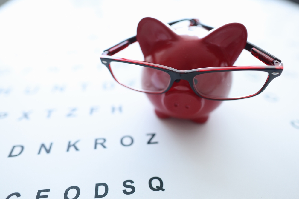
Keeping your eyes healthy is critical for your day-to-day safety as you maneuver around, interact with others, and communicate. Millions of Americans are living with visual impairment, and even more are susceptible for preventable eye diseases and injuries. Below are six things you should be doing now to maintain healthy vision as you age.
Download Maintaining Healthy Vision White Paper
- Reduce Time Spent Staring at a Computer or Tablet: A study from the Vision Council revealed that 68% of millennials suffer from digital eyestrain, which can develop into Computer Vision syndrome (CVS). Technology’s infiltration into our daily activities is unavoidable, but you still need to be mindful of time spent staring at screen by taking 20-minute breaks every couple hours. Dr. Clint Simpson of TLC Eyecare & Laser Centers recommends following the 20-20-20 rule: during those 20 minutes, look at something 20 feet away for at least 20 seconds. Adjusting your workstation so the top of the screen is about 4 to 5 inches below eye level also helps.
- Quit Smoking: Smoking’s consequences are generally associated with cancer and heart disease, but it also affects your vision. The risk of developing age-related cataracts, optic nerve damage and macular degeneration all drastically increase by smoking.
- Take Out Contacts: Leaving your contact lenses in while you sleep deprives your cornea from getting the oxygen it needs from the outside air. Keeping contacts in all the time is like breathing through a mask. Lack of oxygen results in your eyes growing blood vessels where they don’t belong. This has negative effects on your vision. Remember to always wash your hands when placing and removing your contacts and replace them as often as you replace your toothbrush.
- Use Safety Glasses/Goggles: Whether working on a home project, doing yard work, or performing a repair on your car, you need to be protecting your eyes. Dust, debris, chemicals and metal shavings are common things that can get into the eyes and elicit damage. Whenever working on anything that might propel objects into the air, wear safety glasses or goggles.
- Wear Sunglasses: The general rule here is if you are putting on sunscreen, you should be wearing sunglasses as well. Sunglasses will protect your eyes from UV light, which has shown increases in the development of pterygium and cataracts. Make sure the glasses block 99-100% of both kinds of UV light, UVA and UVB.
- See an Eye Doctor: Every 2-3 years you should have a dilated eye exam. After age 45, it should be every 1-2 year(s) in order to screen for glaucoma, cataracts and macular degeneration. For children, a baseline eye screening should be conducted no later than 2 years of age. You should always consult with a professional and not rely on a self-diagnosis with an assist from WebMD. The best way to maintain healthy vision is with regular eye exams.
Protecting your vision starts with simple, everyday habits that make a big difference. From taking regular screen breaks and wearing UV-blocking sunglasses to using proper safety eyewear and scheduling routine eye exams, these proactive steps help you maintain clear, healthy vision for years to come.
Don’t wait until problems arise. Take control of your eye health today and schedule your comprehensive eye exam with Specialty Eye Institute to keep your eyes seeing their best!
RELATED CONTENT: Glaucoma Mini Series

 TLC Eyecare & Laser Centers Retina Institute will be opening its doors this summer!

 877-852-8463