 877-852-8463
 Careers
 Locations
 Patient Portal
 Request Appointment
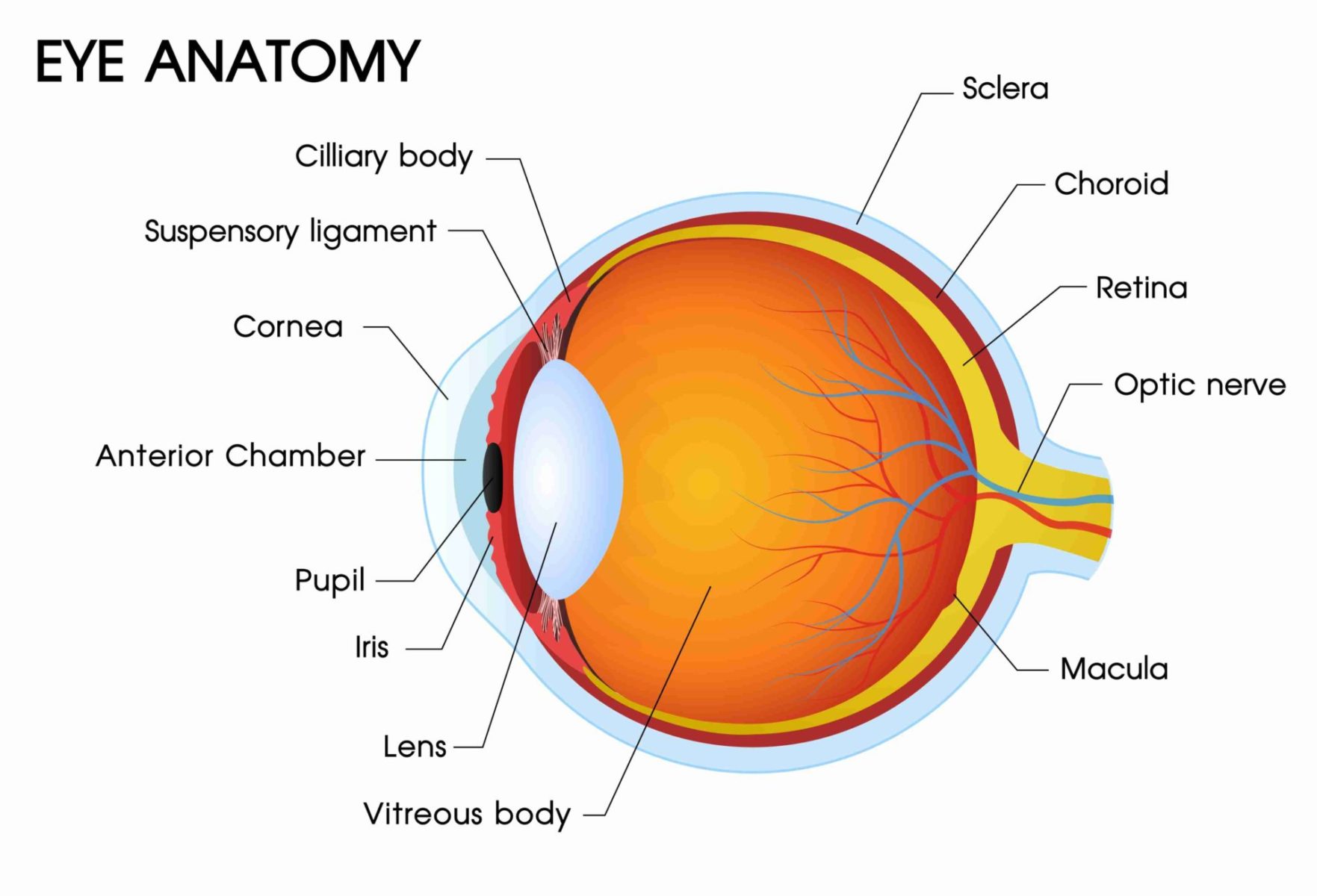
Table of Contents
The Parts of the Eye: Learn the Eye’s Anatomy
The eye is a complex part of your body. It has many different layers and components, each playing a significant role. Learn all about eye anatomy below.
Cornea
The transparent layer forms the front of the eye covers the iris, pupil, and anterior chamber, and provides most of an eye's optical power.
Fovea
The central point in the macula produces the sharpest vision. The fovea contains a high concentration of cones and no retinal blood vessels.
Iris
If you are studying the anatomy of the eye, the iris is the pigmented part of the eye. It is the tissue lying behind the cornea that gives the eye its color (e.g. blue eyes) and controls the amount of light entering the eye by varying the size of the pupillary opening. The iris is the forward extension of the middle (uveal) layer of the eye; separates the anterior chamber from the posterior chamber.
Lens
The natural lens of the eye is a transparent, biconvex intraocular tissue helps bring rays of light to focus on the retina. It is suspended by fine ligaments (zonules) attached between ciliary processes. Think of it like a built-in contact lens!
Macula
Macula is the central area of the retina surrounding the fovea. It is the part of the eye that is responsible for central vision, a.k.a., the largest part of your field of vision!
Optic Nerve
The second cranial nerve. The next component of the eye to be familiar with when you are analyzing eye anatomy is the optic nerve. The largest sensory nerve of the eye; carries impulses for sight from the retina to the brain. It is composed of retinal nerve fibers that exit the eyeball through the optic disc, traverses the orbit, and passes through the optic foramen into the cranial cavity, where they meet fibers from the other optic nerve at the optic chiasm.
Tips & Insights: Learn More About Optic Nerve Damage & Eye Diseases
Pupil
The variable-sized black circular opening in the center of the iris regulates the amount of light that enters the eye.
Retina
Light-sensitive nerve tissue in the eye converts images from the eye's optical system into electrical impulses that are sent along the optic nerve to the brain, to interpret as vision. The retina forms a thin membranous lining of the rear two-thirds of the globe; consists of layers that include rods and cones; bipolar, amacrine, ganglion, horizontal, and Muller cells; and all interconnection nerve fibers.
Vitreous Gel
Transparent, colorless gelatinous mass that fills the rear two-thirds of the eyeball, between the lens and the retina.
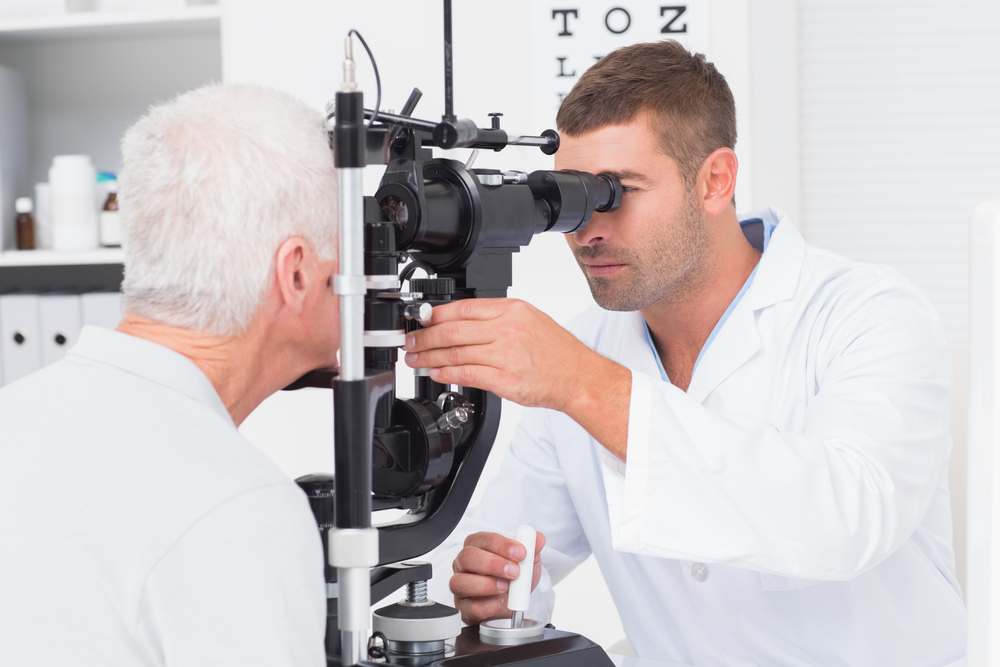
Are Your Eyes Giving You Trouble?
Our team of ophthalmologists offers reliable eye care solutions such as cataract eye surgery, blepharoplasty treatments, Visian ICL eye surgery, refractive lens exchange options, and diabetic retinopathy treatments. Improve your lifestyle with a clear and reliable vision. Give our team of ophthalmologists and optometrists a call by phone at (877) 852-8463 to receive a diagnosis and treatment options for your vision issues.


 877-852-8463