 877-852-8463
 Careers
 Locations
 Patient Portal
 Request Appointment
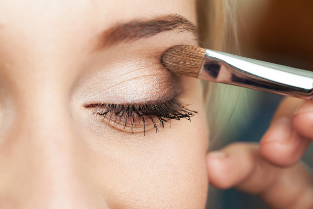
Eye Irritation From Mascara and Makeup Use
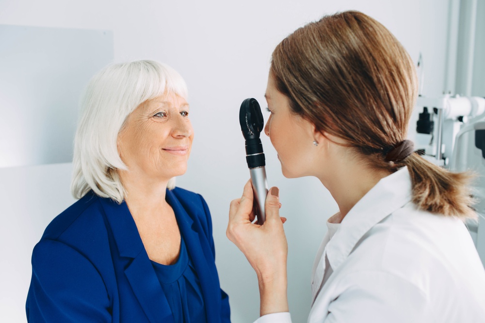
Here’s a question we get asked pretty frequently: Is my eye irritation from makeup use? As eye doctors, it’s something important to consider for any of our patients that use eye makeup and mascara products.
In short, it’s unlikely that you’ll experience vision problems from using eye makeup – but it is possible. Let’s outline the basics.
How Can Eye Makeup Hurt My Eyes?
Since everyone’s eye needs and makeup use routines are different, there’s a lot to be aware of. It boils down to being careful, being clean, and being safe. When it comes to your eyes, you only get one pair, so make sure you take care of them if you use eye makeup!
Below are the top things our eye doctors think you should be careful about.
 Irritation and Infection From Contacts
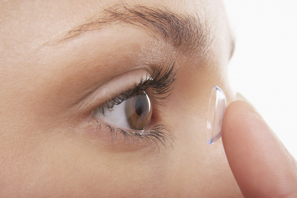
If you wear contacts, your mascara or eye makeup can get into your contacts and ruin the integrity of the lens. This can cause an infection. Make sure you put your contacts in before applying mascara or eye makeup. Be extra aware of the sterility of your hands and lenses.
Scratched Cornea During Application
It’s serious and it’s all too common – scratching the corneas happens when people are not careful when applying mascara. Scratching a cornea can lead to dangerous infections, so make sure to be careful when applying mascara or makeup.
Try to avoid applying eye makeup or mascara in the car – that’s one of the most common places where people will scratch a cornea.
Avoid using mascara that uses fibers to thicken and lengthen the lashes. The fibers may fall into the eye, scratching the cornea.
Tips & Insights: The Benefits of Receiving LASIK Eye Surgery
Allergic Reactions to Eye Makeup
If you’re sensitive to certain materials or are known to have allergic reactions, you should always test makeup before putting it on to avoid eye irritation. Some brands will label their makeup as hypoallergenic or ophthalmologist-approved. If you’re having allergic reactions in your eyes to makeup, you should talk to an eye specialist about your options.
Sleeping in Mascara
Some makeups have non-toxic components, like glitter, that could cause irritation, especially if it gets into your eyes while you’re sleeping.
We cannot stress this enough – remove your makeup at the end of the night before you sleep. Try not to fall asleep with any makeup on – especially anything near your eyes.
Eye Irritation From Expired Makeup
A lot of people tend to ignore the expiration dates on their makeup and mascara – but the reason that they’re in place is that makeup tends to have preservatives in it to keep bacteria from building up inside of it. The preservatives do not work after the expiration date. Do not keep expired makeup. The eye irritation is not worth it!
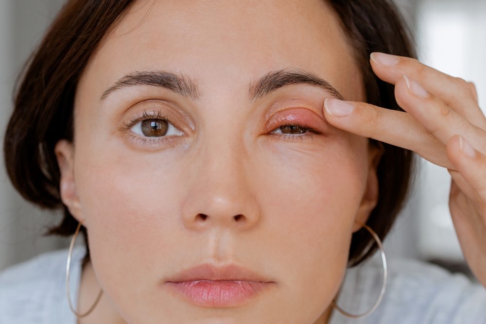
Can Mascara Cause Pink Eye?
If you share your makeup with others or don’t clean your makeup brushes, bacteria can grow. This can cause a bacterial infection, such as conjunctivitis, more commonly known as pink eye.
Make sure not to share your makeup, especially with people who have been around others with bacterial infections.
Also, make sure to properly conceal your makeup when you’re done using it. If you’re using dry makeup, you may be able to sanitize it with alcohol wipes. Be aware that some makeup may dissolve in alcohol.
Caring For Your Vision With Specialty Eye Institute
Our team of ophthalmologists and doctors is committed to preserving and maintaining your vision. Our doctors offer a variety of vision care treatments such as cataract surgery, blepharoplasty treatments, corneal transplants, presbyopia treatments, and LASIK eye surgery. Give our team a call by phone at (877) 852-8463 or book an appointment online to receive support. Our staff will help you choose surgery or treatment that aligns with your issues.
Tips & Insights: Available Cataract Surgery & Treatment Options


 higan’s Best and Brightest Companies to work for the second consecutive year in a row. SEI was recognized for demonstrating exceptional innovative human resource practices.
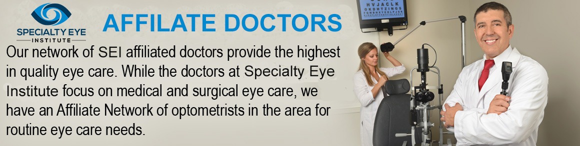
 877-852-8463