 877-852-8463
 Careers
 Locations
 Patient Portal
 Request Appointment
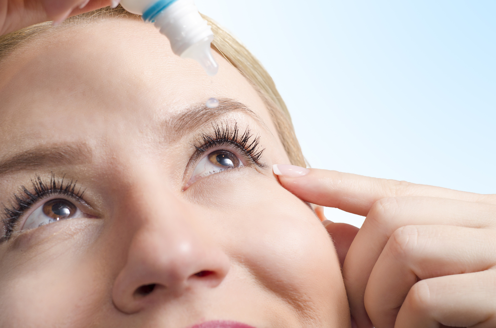
 This year more than ever, a teacher's job can be difficult, especially when glasses or contacts get in the way. That’s why we would like to show our support for our local teachers and make their jobs and lives a little bit easier.
Teachers can take advantage of $500* OFF LASIK! Schedule your FREE LASIK Consultation by filling out the form below.
*$500 off discount ($250 off per eye) is applicable towards bilateral LASIK or PRK. Cannot be combined with other offers or insurance.


 877-852-8463